
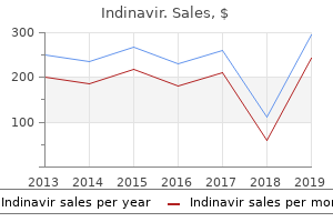
Indinavir
"Buy indinavir 400mg with visa, medicine x xtreme pastillas".
By: H. Merdarion, M.B. B.CH. B.A.O., M.B.B.Ch., Ph.D.
Clinical Director, Medical College of Wisconsin
There is support in modern era texts medications that cause hair loss order discount indinavir line, concluding that the use of radiation "may provide an alternative to conventional conservative treatment for patients who are not surgical candidates" (PerezBrady) treatment keratosis pilaris order indinavir 400 mg without prescription. Typical treatment is with photon beam therapy using treatment 7th feb bournemouth purchase generic indinavir on-line, at most medications affected by grapefruit purchase 400mg indinavir with mastercard, complex treatment planning in five or fewer fractions. The presentation and behavior ranges from truly benign to aggressive with metastatic potential. Surgical resection has historically been the treatment of choice with radiation reserved for technically or medically inoperable cases. Precise histologic classification may help discriminate those truly benign lesions that would not be expected to benefit from radiation therapy from lesions that would be best treated as invasive carcinomas. For those unresectable non-secretory lesions causing symptoms such as pain, radiation may be beneficial. For secreting tumors, radiation therapy is limited to those causing symptoms that are not controllable by medical means. The relationship to subsequent malignant lymphoma is unclear, with malignant lymphoma reported in as many as 30% of cases. Synonyms include giant follicular lymph node hyperplasia, follicular lymphoreticuloma, angiomatous lymphoid hamartoma, and giant benign lymphoma. Low dose radiation therapy has been reported as effective in refractory or relapsed cases if further use of steroids is contraindicated. Castration There is evidence that with sufficient dose radiation can effectively and permanently cease gamete production and hormone production in the testes and ovaries. Surveys reported by Order and Donaldson (1998) indicated 75% of surveyed radiation oncologists would use radiation for this purpose with the appropriate indication. Department of Health, Education, and Welfare survey report of 1977 included castration as an acceptable indication. The availability of drugs which achieve the same result has largely rendered this as obsolete. Chemodectoma (carotid body, glomus jugulare, aortic body, glomus vagale, glomus tympanicum) (chromaffin negative) Chemodectoma is a general term that includes many specific types based on the location of the body in which they arise. These are chromaffin-negative, benign tumors that can arise in the chemoreceptor system, such as the aortic body; carotid body; glomus jugulare; and tympanic body. It is generally accepted that radiation therapy, with or without surgical resection, is medically necessary, with a significant probability of control. These tumors of notochord origin can be benign or malignant, but all tend to be locally invasive and tend to recur locally, some with the potential to metastasize. Surgery is the primary approach, but is often inadequate to control the primary tumor. Postoperative radiation therapy, and radiation therapy for inoperable lesions, is considered medically necessary. Adjuvant radiation is not indicated unless there is progression that cannot be dealt with surgically. Choroidal Hemangioma these are rare vascular tumors and may be circumscribed or diffuse, the latter associated with Sturge-Weber syndrome. Typically, radiation therapy is given using complex or three dimensional conformal external photon beam technique, or using low dose rate brachytherapy plaque. Corneal Vascularization Radiation therapy is not indicated in the treatment of corneal neovascularization. Corneal xanthogranuloma Corneal xanthogranulomas may develop in association with generalized juvenile xanthogranuloma and generalized histiocytosis. Reports in old literature of the treatment by contact radiation or photons do not establish any definite benefit. First line therapy, when observation is not selected, is steroid therapy or surgery. Craniopharyngioma Most often radiation therapy is used as an adjuvant after maximal safe resection. Local control rates are similar whether radiation is given at time of first relapse or immediately after surgery. Dermatitis Skin inflammation from a variety of etiologies (both known and unknown) has been treated in the past by using low dose, very superficial radiation or Grenz rays. The use of radiation for this purpose is reserved for cases refractory to non-radiation measures.
However symptoms yeast infection men discount indinavir online master card, in the presence of competing risks medications covered by blue cross blue shield discount indinavir 400 mg without a prescription, the other causes of death are handled in a different manner symptoms 5 days after conception cheap 400 mg indinavir free shipping. Under such circumstances symptoms nausea indinavir 400 mg otc, it is not possible to compute a cause-adjusted survival rate. However, it is possible to adjust partially for differences in the risk of dying from causes other than the disease under study. This can be done by means of the relative survival rate, which is the ratio of the observed survival rate to the expected rate for a group of people in the general population similar to the patient group with respect to race, sex, and age. The relative survival rate is calculated using a procedure described by Ederer et al. It is always greater than the observed survival rate for the same group of patients. The existence of true population values is postulated, and these values are estimated from the group under study, which is only a sample of the larger population. The difference between the two results is called the sampling variation (chance variation or sampling error). The standard error is a measure of the extent to which sampling variation influences the computed survival rate. In repeated observations under the same conditions, the true or population survival rate will lie within the range of two standard errors on either side of the computed rate approximately 95 times in 100. For example, the starting time for studying the natural history of a particular cancer might be defined in reference to the appearance of the first symptom. Various reference dates are commonly used as starting times for evaluating the effects of therapy. These include (1) date of diagnosis, (2) date of first visit to physician or clinic, (3) date of hospital admission, (4) date of treatment initiation, date of randomization in a clinical trial evaluating treatment efficacy, and (5) others. The essential question is, "What is the probability that the observed difference may have occurred by chance If the 95% confidence intervals of two survival rates do not overlap, the observed difference would customarily be considered statistically significant, that is, unlikely to be due to chance. This latter statement is generally true, although it is possible for a formal statistical test to yield a significant difference even with overlapping confidence intervals. Moreover, comparisons at any single time point must be made with care; if a specific time (5 years, for example) is known to be of interest when the study is planned, such a comparison may be valid; however, identification of a time based on inspection of the curves and selection of the widest difference make any formal assessment of difference invalid. It is possible that the differences between two groups at each comparable time of follow-up do not differ significantly but that when the survival curves are considered in their entirety, the individual insignificant differences combine to yield a significantly different pattern of survival. The most common statistical test that examines the whole pattern of differences between survival curves is the log rank test. This test equally weights the effects of differences occurring throughout the follow-up and is the appropriate choice for most situations. Other tests weight the differences according to the numbers of persons at risk at different points and can yield different results depending on whether deaths tend more to occur early or later in the follow-up. Care must be exercised in the interpretation of tests of statistical significance. For example, if differences exist in the patient and disease characteristics of two treatment groups, a statistically significant difference in survival results may primarily reflect differences between the two patient series, rather than differences in efficacy of the treatment regimens. The more definitive approach to therapy evaluation requires a randomized clinical trial that helps to ensure comparability of the patient characteristics and the disease characteristics of the two treatment groups. At any given time, the vital status of each patient is defined as alive, dead, or unknown. In each case, the observed follow-up time is the time from the starting point to the terminal event, to the end of the study, or to the date of last observation. This observed follow-up may be further described in terms of patient status at the endpoint, such as the following: Alive; tumor-free; no recurrence Alive; tumor-free; after recurrence Alive with persistent, recurrent, or metastatic disease Alive with primary tumor Dead; tumor-free Dead; with cancer (primary, recurrent, or metastatic disease) Dead; postoperative Unknown; lost to follow-up Completeness of the follow-up is crucial in any study of survival, because even a small number of patients lost to follow-up may lead to inaccurate or biased results. The maximum possible effect of bias from patients lost to follow-up may be ascertained by calculating a maximum survival rate, assuming that all lost patients lived to the end of the study.
Generic 400mg indinavir free shipping. He Killed His Family BECAUSE OF PUBG -19-year-old Delhi boy stabs family 33 times.
The regional lymph nodes draining the bladder include primary and secondary nodal drainage regions medications 5 rights buy 400 mg indinavir. The presacral lymph nodes are classified as a primary drainage region; however medications used to treat depression order indinavir cheap online, mapping studies have found this area to be a less frequent site of primary regional metastases medicine in french buy indinavir amex. Primary nodal regions drain into the common iliac nodes symptoms joint pain and tiredness buy generic indinavir 400mg on-line, which constitute a secondary drainage region. Regional lymph node staging is of significant prognostic importance given the negative impact on recurrence after treatment and long-term survival. The relevant information from regional lymph node staging is obtained from the extent of disease within the nodes (number of positive nodes, extranodal extension) not in whether metastases are unilateral or contralateral. Pathologic staging is based on the histologic review of the radical or partial cystectomy specimen. Total cystectomy and lymph node dissection generally are required for this staging; however, a pathologic staging classification should be given for partial cystectomy specimens. Pathologic staging should include the findings of the cystectomy specimen following surgery and should be assigned independent of previous clinical or biopsy information that is used for clinical stage assignment. Adequate nodal staging requires removal of the primary lymph node regions that include the left and right external iliac, hypogastric and obturator nodes. Based on contemporary mapping studies in which standard techniques were used to evaluate the pathologic specimen, excision of the primary nodal regions should result in an average of >12 lymph nodes. Evaluation of the National Cancer Database revealed a significant difference in survival in those patients who had fewer than four lymph nodes removed compared with those who had more than four lymph nodes removed, even in patients with node negative (N0) disease. This should serve as a guide for the num- ber of lymph nodes to be evaluated for optimized staging after radical cystectomy. However, the lymph nodes examined may vary dependent on previous patient treatment, body habitus, and pathologic technique. The number of lymph nodes examined from the operative specimen and the number of positive lymph nodes have been reported to be associated with survival. In addition, the size of the largest tumor deposit and presence of extranodal extension may independently impact survival. Primary tumor stage and grade are important independent predictors of tumor progression and outcome. More recently morphologic prognostic features including lymphovascular invasion and variants of the pattern of tumor growth, such as micropapillary and nested variants, have been found to portend an adverse outcome. Lymph node status has a profound effect on the risk of tumor recurrence and patient survival. Various Urinary Bladder 499 In order to view this proof accurately, the Overprint Preview Option must be set to Always in Acrobat Professional or Adobe Reader. Job Name: - /381449t lymph node parameters demonstrating prognostic significance include the total number of excised lymph nodes, the number of positive lymph nodes, extranodal tumor extension, and the ratio of number of positive lymph nodes to total number of lymph nodes evaluated. Several molecular factors with prognostic importance have been identified for bladder cancer. These markers are involved in the regulation of the cell cycle, programmed cell death, growth factor signaling, and angiogenesis. Two distinct molecular pathways for bladder tumor progression have been established. Noninvasive tumors appear to progress through a pathway that involves the frequent alteration to chromosome 9, specifically 9q deletions. In contrast high-grade tumors are associated with a loss of heterozygosity of chromosome 17p, 14q, 5q, 3p. In the setting of advanced disease, patient performance status, the presence of visceral metastases, and elevated levels of alkaline phosphatase are important predictors of response to systemic therapy and patient survival. Regional Lymph Nodes (N) Regional lymph nodes include both primary and secondary drainage regions.
As multiple primary formations Siedamgrotzky once counted in a thirteen-yearold stallion more than a hundred fibromata in the skin treatment writing purchase indinavir 400mg fast delivery, which had pedunculated grown in the course of three months in the breast medications prescribed for pain are termed buy genuine indinavir online, a abdomen treatment trichomoniasis cheap 400 mg indinavir visa, neck and face treatment table discount indinavir 400 mg with visa, to sizes varying from that of hazel nut to that of the palm of the hand. Fibromata are frequently found in the tongue of the cow, as well as in the vagina, in the latter situation as pedunculated growths of large size projecting into the vaginal Those fibromata which are found in horses and cattle canal. Fibromyxomata are especially common at the end of the tail and in the heart (epicardium and endocardium) as pedunculated growths, as well as in the naso-pharnyx (where they are found hanging from the wall; covered with mucous membrane and are known as nasal polyps). The author has observed be harmful because of its size and location (occlusion of passages, pressure upon vital organs), but does not give rise to metastasis and usually does not recur after fibroma extirpation. A may sometimes the hard fibromata of the skin and tongue undoubtedly are often results of traumatic lesions. The lipoma clusters or fatty tumor cells is made up stages of of adipose tissue, of of fat in various development, along with bloodvessels and fibrous connective tissue. The peritoneal adipose tissue often forms lobular appendages, which when much overgrown and stretched movements become pedunculated and present pearshaped masses or tumors of the size of a fist. Lobulated lipomata are in the intes- found also tine cow and hog tissue is in the omentum, and is and perirenal fat; in these animals the fat firm and white. The subcutaneous in the size a frequent site in the horse, especially neighborhood of the knees where they may a attain very large (twenty-six; and a half kilograms in case reported by Moller) and in the dog especially on the inner surface of the thigh, in the fold of the knee (Frohner), and in the breast (Stockfleth). According to Bostrom it pos- a bit of germinal fat tissue of the skin of the embryo might have been separated and been misplaced in the cranial Lubarsch suggests that intraparenchymatous renal lipoarea. It is characterized microscopically by an intercellular substance rich in mucin, in which are sparsely scattered cells of spindle and highly branched stellate forms with long,: 4j s. In fresh material the mucinous substance may be precipitated by the addition of acetic acid. Often only individual portions of the tumor are conspicuously myxomatous, the remainder being made up of fat tissue or dense fibrous tissue (myxolipoma, jiiy. Myxomata develop as single or growths according to their more or less multiple primary situation, and as sarcomatous mixed tumors capable of metastasis and of embryonic appearance. They are especially met upon and in the heart of cattle (when the subpericardial and subendocardial foetal mucous tissue acts as their developmental substance) as rounded, lobulated growths ranging in that of a size; from that of a nut to fist they also occur in the nose in the cow and horse, in the parotid, and in in one case in the dog along the nerve around the spinal cord (Holzmann), and. A chondroma or cartilaginous tumor connective tissue has, in addition its to a main constituent cartilaginous tissue, usually of the hyaline type, and contains cartilaginous cells of very irregular size encapsulated in the fibrous this substance without regularity of arrangement. Besides the pure chondromata there are met many mixed types, made up of fibrous tissue, bone and glandular tissue, in which cartilage enters as only one of the constituents and modifications may also result from the rather marked tendency of the cartilage to metamorphosis, both regressive and progressive. Muvascular fibrous framework, as; coid degeneration with production giving of gelatinous to softened foci (cysts), and calcification, rise white opaque patches Chondroma. Chondromata are nodular, nodose, lobulated, rounded tumors of dense (in case of myxomatous change or when other tissues are combined they are softer) consistence showing their cartilaginous substance as a milk-white, grayish and bluish white, semitransparent material. They may attain considerable dimensions, perhaps the size of a human head, and may weigh from ten to twenty-eight kilograms. According to their location the cartilaginous tumors may arise from previously existing cartilage (ec chondromata) or may develop within the bone marrow or in;, the soft tissues which do not contain cartilage (enchondromata) in the former probably originate from the perichondrium, the cells of which are usually concerned from formation of cartilage; the latter arise misplaced embryonic cartilaginous rudiments. Isolations of bits of tissue from either of these sources in may occur embryonic life from developmental faults, as in the formation of the branchial arches or the primitive vertebrae and embryonal ital elements of the ribs. Chondromata are most common in the bony framework; of the chest as the result of fractures of the ribs, projecting into the chest cavity or beneath the skin as large also frequently met in the gland, originating from remnants of the branchial cartilages, in goats, horses, dogs and cattle. As mixed growths (chondroadenoma) they have been seen a number of times in the lungs, mammary gland and in the testicles in animals. As exceptionally rare instances may be mentioned enchondromata of the vitreous tumor masses they are neighborhood of the jaw and thyroid Osteoma. A chondroma should be looked upon pening to penetrate by growth into the veins sometimes develop long processes within the lumen of the vessels, which are of course obstructed and any little portions which may be carried away by the blood may form metastatic nodes in other organs. Where the bone trabecula have no lamellated structure and the bone corpuscles do not show the usual projecting branches the specimen is spoken of as an osteoid sarcoma. The flat small protuberances, of the size of a pea to that of a nut, projecting from the surface of bones, are also called exostoses; the superficial thickenings around bone shafts, periostoses; those which grow along bones as bony formations in fascise and connective tissue, parostoses; an ivory-like, hard, well-defined formation in the interior of Diffuse thickenings of the bones in the bones, an enostosis. This view is especially applicable to the rather common large osteomata of the head cavities (cattle, horses), arising from the sphenoid, ethmoid or the turbinate bones in their gradual enlargement they press upon the surrounding bony walls, force the jaw and nose out of position, project into the maxillary sinus, nasal cavities or into the orbit or cranial cavity, and obstruct these spaces with continuous pressure upon the respective soft parts.